Thursday, March 1, 2012

heart dissaction


I used the sagittal cut the to separate into two pieces.
Reflection Questions:

1. After viewing the artery and vein prepared slide, which blood vessel has the thickest wall? Is there a possible reason for this, on the basis vessel function? The artery is the thickest wall, and the reason why it is possible is because of the amount of blood it has to carry.

2. After viewing the cardiac muscle prepared slide, is cardiac muscle smooth or striated? Cardiac muscle is smooth not shriated

3. The prepared slide of a coronary artery with atherosclerosis, what is the danger of having the atherosclerotic plaque on this particular artery? the dangers is that it will lock the blood flow.

4. How does the basic structure of the heart compare between the three heart specimens? The heart closest reasonable the pig because of similarity between them but all in all most of hearts have similar structures.

5. What are some o  the major differences you observe in the heart specimens?  the smaller parts are different from each other but basically the same except for smaller details.

6. Can you think of any adaptive reason for these differences? Animals hearts are more adapted to fit their lifestyle and also have longer arteries to pump blood faster while the human hearts are smaller and compacted to fix our size.

Thursday, February 9, 2012

Hearing Project*

leech lab*/ Neurophysiology

Objective:  
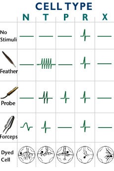
This lab was to identify,analyze a neuron. Also was to record electrical activities of individual neurons while we delivered mechanical stimulus to the attached skin, inject fluorescent dyes into the neurons to visualize their morphology, and identify the neurons based on the morphology and the response to stimuli, comparing them to previously published results. This was also to help us to learn neurophysiology- study of physical and chemical processes of neurons.


Materials:
leech
scalpel
pins
dissection tray
probe
forceps
feather
scissors
leech tank
20% Ethanol
tongs
dissection microscope
micro-manipulator
Oscilloscope


Steps:
Step 1
Catch and anesthetize the leech in 20% ethanol solution. Ethanol is not an anesthetic for vertebrate animals, but can be an effective anesthesia for small creatures that breathe through the skin like the leech. Like in many things, too high a concentration will be harmful or fatal.

Step 2
Pin the animal dorsal side up through the anterior and posterior suckers onto a dissection tray, stretching the animal in the process.

Step 3
Using scissors, make a cut in the skin along the mid-line on the dorsal surface, taking care not to damage deep structures. Using forceps, carefully tease apart the skin along the cut and pin down the left and right halves of the skin to each side, so that the leech is pinned open with the inside of the skin facing up. This exposes the innards of the leech, including the digestive, excretory and reproductive organs. You cannot see the nervous system yet, because they are located ventrally.

Step 4
Carefully remove the gut and other internal structures to expose the ventrally located nerve cord. The nervous system of the leech is encased within the ventral sinus, which is dark green in color.

Step 5 
Notice that there are many swellings up and down the sinus. These contain the segmental ganglia of the nervous system. To make one of them accessible, first we cut a window in the body wall underneath a ganglion, taking care not to damage the nerve cord or any attached nerves in the process.


Step 6
Isolate a section of the animal by making 2 parallel cuts across the animal (perpendicular to the anterior-posterior axis), but sufficently separated so that the strip you remove contains at least one ganglion. Then, with forceps, flip the piece of skin over so that the outer skin is now face up. Pin the skin down


Step 7
Cut the sinus with an ultra fine scalpel and using fine forceps, carefully tease apart the sinus to expose the ganglion. Individual cells can now be viewed under the microscope. In reality, you would only use the scalpel here only if you are extremely good at microdissection. It's very difficult to cut just the sinus without accidentally damaging the ganglion underneath, but hey, we are all perfect in cyberland. Normally, this is done with a pair of very fine forceps.

Step 8
Now you've come to the crux of the matter. All the preparation so far has been to make this step possible. You might want to review Nervous System background or Electrical Equipment background at this point. Click on the electrode to gain control of it. Move the electrode to somewhere over the ganglion then click on the mouse button. This simulates the process of penetrating the cell, which is much more demanding in reality. Keep your eyes glued to the oscilloscope display while you are doing this. If you find a cell, the display will change. If you see no change, then you have not found a cell. Keep moving your electrode around and clicking until you find a cell. The sound you hear is the oscilloscope display you are seeing fed into an audio amplifier. It provides an audio feedback to what you see on the screen. Now using a feather, probe or forceps, push around the skin of the animal. Observe if the cell you have penetrated responds to weak (feather), medium (probe), strong (forceps) or any stimulus. Note the pattern of response. The cell may fire action potentials or spikes. The response characteristics will be used when you are comparing your data with published data compiled in the atlas. When you are satisfied with the electrophysiology, you can start the anatomical investigation by injecting the cell with a fluorescent dye. Push the button labeled "Dye Injection."

Step 9
Next, we will visualize the morphology of the neuron from which you have just recorded using a fluorescent dye. Having pushed the button labeled "Dye Injection," the amplifier system has passed an electric current from the electrode that resulted in the ejection of Lucifer Yellow from the tip of the electrode into the intracellular space. Lucifer Yellow will passively spread throughout the cell after a while. Now you can turn on ultraviolet (UV) light by pushing "UV Switch.". Lucifer Yellow fluoresces bright yellow-green under UV and you will be able to visualize the cell in question, including its axon, dendrites, cell body and so on.

Step 10
You now have electrophysiological data and neuroanatomical data from your experiment. Try to identify the cell based on published data (Atlas) There are many cells in different locations of this ganglion. Repeat the whole procedure for as many cells as you would like.


This lab taught me how to identify the different cell types and how the cells  countered to different stimuli.

http://www.hhmi.org/biointeractive/vlabs/neurophysiology/index2startlab.html

Friday, January 20, 2012

neuron*

A neuron is a nerve cell that is specialized to carry messages through the body by transmitting signals to the nervous system.There are 100 billion neurons in our nervous system. Neurons come in all different shapes and size, the biggest neuron is 3 meters long and the smallest is only 4 microns wide. Neuron are very similar to the other cells in the body, as that may be they actually are very different. The neurons contact with each other by using the electrochemical process. Also neurons have elaborated with dendrites and axons. The dendrites deliver info to the cell and look like spikes coming out of the cell body. And the axons take the information from the cell body and are hard to classify from the dendrites. The axon conduct electrical messages that is called action potentials.  Normally when the neuron is at rest the positive charges are outside and the negative charges are inside of the axon of a cell and it's not not sending a signal which is resting potential. When its at rest the potassium ions cross through the membrane, but the chloride ions and sodium ions have a difficult time going through. A resting membrane potential of the neuron is 70mv. There are relatively more sodium ions outside the neuron and more potassium ions inside that neuron. But then when it increases to about 50mv its is called action potential. 


http://faculty.washington.edu/chudler/ap.html
http://faculty.washington.edu/chudler/cells.html
http://webspace.ship.edu/cgboer/theneuron.html

Friday, December 16, 2011

Joints!


There are three structural classification in the joint family. They are fibrous, cartilaginous, and synovial. They blind joints together. There are also three other functional classes of the joints that are on the amount of movement they have. The first is synarthroses, which is immovable. The second is amphiarthroses, and it is slightly movable. The third is diarthroses, and that is able to move freely. There is also fibrous structural joints, which means that bones are joined and filled by fibrous tissue. And there is no joint cavity. These bones are very tightly bind together. One of the types of fibrous structural joints is syndsmose. This is connected with tibia ad fibula and movement mostly in immovable. Then there are synovial joints. Synovial joints have articular cartilage, synovial fluid, and reinforcing ligaments. These joints are bones that are separated by fluid. It is located the limb joints. There are two muscle attached across the joints, they are origin that are immovable and insertion that can be able to move. Arthritis is one of the most common it joint problems. There are more than 100 different types of diseases that damage the joints. People that getting older tend to get it easier because the cartilage gets destroyed faster. The end of the bone thickens and enlargers which causes the bone to have spurs and painful to move. This can effect a lot of parts in you body such as: knuckles, knees, and spins.

Monday, November 7, 2011

Skin*

SKIN:


Skin is composed of different tissues that helps protect from sun rays that could enter the body. And it also helps regulate the body temp, and it restores water loss. Skin has vitamin D, that help the immune system and waste system. There are 2 layers, the first is the epidermis,squamous epithelium then the dermis, thicker and made out of elastic fibers. Collagen is inside the skin which help smooth the muscle tissue, nervous tissue, and the blood. There are types skin cancers and burns that can damage the skin.


SKIN CANCERS:

There are 1 million cases a year and it kept increase more and more each year.  There are three different types of skin cancer from the least to the more dangerous stages:Basel cell carcinoma,Squamous cell carcinoma,Melanoma. Skin cancer in most common in light skinned people that are usually outside a lot. Skin cancer has many different looks like flat or raised, dry, hard and different colors like black or brown.It is mostly found in the neck scalp or face. There are many ways to prevent from getting skin cancer.  Limiting recreational sun exposure, avoiding unprotected exposure to the sun during peak radiation times, wearing hats and tightly protecting clothing while outdoors. Also regularly using a waterproof sunscreen with UVA protection and SPF 30 or higher, and avoiding the use of tanning beds.


BURNS:


Common burns would be the minor sun burns that mostly everyone tends to get. A 1st degree burn injures the epidermis. It will heal most likely in a week or so without scaring. A 2nd degree burn damages the epidermis and the deep partial thinness. You get this burns from hot objects and it heals without scaring if it is not infected. And a 3rd degree destroys the epidermis, dermis, and structures of the skin. This cause skin the become very dry. This is cause by hot object, chemicals, flame, hot liquid. And it heals only if there is a growth epithelial cells.